Label Diagram of Ear
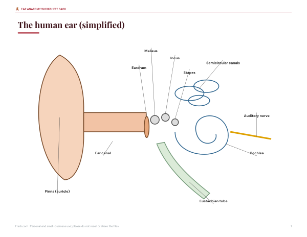
Explore a labelled diagram of the ear, then test yourself. Click any label to learn what that part does, hide the labels to quiz yourself, and follow how sound travels from the outer ear to the brain. The diagram is simplified for learning — perfect for biology homework, revision and teaching — and printable labelled and blank worksheets are included in the pack.
- Free to use
- No sign-up
- Runs in your browser
Ear diagram and labelling quiz
Ear Anatomy Worksheet Pack
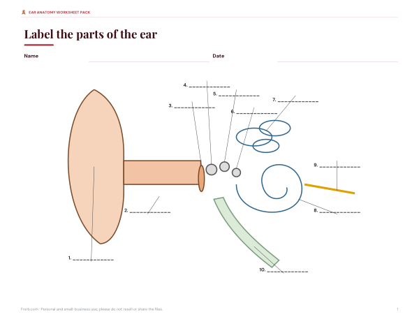
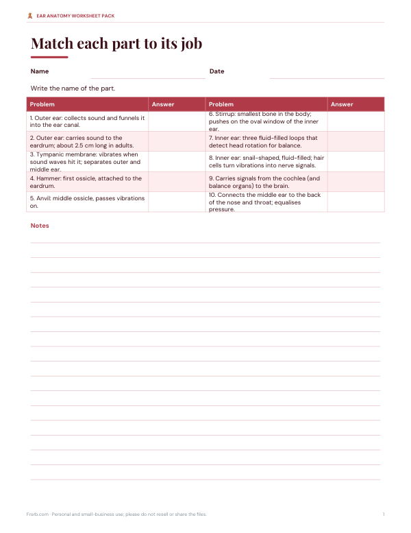
Printable labelled ear diagram, a blank labelling worksheet with answer key, a parts-and-functions matching worksheet and a “path of sound” sequencing sheet.
- Labelled diagram (PDF)
- Blank worksheet (PDF)
- Parts and functions (PDF, DOCX)
- Path of sound (PDF/DOCX)
Formats: PDF, DOCX. Instant download after payment (link valid 72 hours, up to 5 downloads). AI-assisted: the templates were drafted with AI help and reviewed and laid out before publishing.
$5.00 USD, one-time 4 files · about $1.25 each
- Instant download
- 7-day refund
- Secure checkout by Stripe
7-day money-back guarantee. Full refund within 7 days of purchase, even after you download.
Free sample: Blank Ear Diagram Worksheet (PDF, 59 KB)
Full refund within 7 days — see the refund policy and license.
Free printables
Single sheets you can print right now, no sign-up. The full pack adds every sheet in one download plus the editable files.
 Printable Ear Sound Path Worksheet
The three parts of the ear
| Region | Structures | Main job |
|---|---|---|
| Outer ear | Pinna (auricle), ear canal | Collects sound and channels it to the eardrum |
| Middle ear | Eardrum, malleus, incus, stapes, Eustachian tube | Turns sound waves into vibrations and amplifies them |
| Inner ear | Cochlea, semicircular canals, vestibule, auditory nerve | Turns vibrations into nerve signals; senses balance |
How we hear: step by step
- Sound waves are collected by the pinna and travel down the ear canal.
- They make the eardrum vibrate.
- The three ossicles — malleus, incus and stapes — pass the vibrations along and amplify them.
- The stapes pushes on the oval window, setting the fluid inside the cochlea moving.
- Tiny hair cells in the cochlea bend with the fluid waves and produce electrical signals.
- The auditory nerve carries the signals to the brain, which interprets them as sound.
Different parts of the cochlea respond to different pitches: high-pitched sounds near its base and low-pitched sounds near its tip.
Parts of the ear explained
| Part | What it does |
|---|---|
| Pinna (auricle) | Outer ear: collects sound and funnels it into the ear canal. |
| Ear canal | Outer ear: carries sound to the eardrum; about 2.5 cm long in adults. |
| Eardrum | Tympanic membrane: vibrates when sound waves hit it; separates outer and middle ear. |
| Malleus | Hammer: first ossicle, attached to the eardrum. |
| Incus | Anvil: middle ossicle, passes vibrations on. |
| Stapes | Stirrup: smallest bone in the body; pushes on the oval window of the inner ear. |
| Semicircular canals | Inner ear: three fluid-filled loops that detect head rotation for balance. |
| Cochlea | Inner ear: snail-shaped, fluid-filled; hair cells turn vibrations into nerve signals. |
| Auditory nerve | Carries signals from the cochlea (and balance organs) to the brain. |
| Eustachian tube | Connects the middle ear to the back of the nose and throat; equalises pressure. |
Balance: the vestibular system
The inner ear also keeps you balanced. The three semicircular canals sit at right angles to each other and contain fluid that moves when you turn your head; sensory hair cells detect this movement. The vestibule, between the canals and the cochlea, contains organs that sense gravity and straight-line acceleration. Together they send information to the brain about head position and movement — which is why inner-ear problems can cause dizziness.
The Eustachian tube and ear pressure
The Eustachian tube links the middle ear to the back of the nose and throat. It opens when you swallow or yawn, letting air in or out so pressure on both sides of the eardrum stays equal. On a plane or in a lift, a blocked tube can make ears feel full or painful until it opens — swallowing, yawning or chewing usually helps. Colds can block the tube and contribute to middle-ear infections, especially in children, whose tubes are shorter and more horizontal.
Using the diagram to study
- Start with labels shown and click each part to read its role.
- Say the path of sound aloud: pinna → ear canal → eardrum → malleus → incus → stapes → cochlea → auditory nerve → brain.
- Hide the labels and use the quiz until you can find every part.
- Print the blank worksheet and label it from memory.
- Check with the labelled version and repeat tricky parts.
Common ear problems (for context)
| Condition | Where | What happens |
|---|---|---|
| Earwax build-up | Outer ear | Wax blocks the canal and muffles sound |
| Otitis externa (“swimmer’s ear”) | Outer ear | Infection or inflammation of the ear canal |
| Otitis media | Middle ear | Middle-ear infection, often after a cold |
| Perforated eardrum | Middle ear | A hole in the eardrum from infection or injury |
| Noise-induced hearing loss | Inner ear | Loud sound damages cochlear hair cells |
| Vertigo | Inner ear | Balance organs send false movement signals |
This table is for learning only; anyone with ear pain, discharge, hearing loss or dizziness should seek medical advice.
Protecting your hearing
Hair cells in the cochlea do not regrow once damaged, so noise-induced hearing loss is permanent. Turn down headphones (a common guideline is no more than 60% volume for no more than 60 minutes at a time), wear ear protection at concerts and with loud tools, and give your ears quiet breaks after noisy events.
Memory tricks
- The ossicles go in alphabetical order from the eardrum inwards: Malleus, Incus, Stapes — or remember “hammer, anvil, stirrup”.
- Cochlea looks like a snail shell (cochlea is Latin for snail).
- Semicircular canals are three “loops for balance”.
- The stapes is the smallest bone in the human body — about 3 mm long.
A note on the diagram
The diagram is a simplified schematic designed for learning the parts and their order. Real ear anatomy is three-dimensional and much of the middle and inner ear is enclosed in the temporal bone of the skull. For medical questions about hearing or ear symptoms, see a qualified health professional.
Privacy
The diagram and quiz run in your browser.
Frequently asked questions
What are the three parts of the ear?
The outer ear, middle ear and inner ear.
What are the three smallest bones in the ear?
The malleus, incus and stapes, called the ossicles.
What does the cochlea do?
It turns vibrations into nerve signals for hearing.
Which part of the ear controls balance?
The semicircular canals and vestibule in the inner ear.
What is the Eustachian tube for?
It equalises air pressure between the middle ear and the throat.
Can I print the diagram?
Printable labelled and blank worksheets are in the paid pack; the interactive diagram is free.
What is the smallest bone in the body?
The stapes in the middle ear, about 3 mm long.